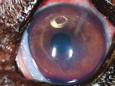
PIGMENTED DEGENERATION / atrophy / progressive retinal (PRA PROGRESSIVE Retinal Atrophy) IN THE DOG AND CAT IN THE DEEP CORNEAL ULCER
progressive retinal atrophy and 'disease on a hereditary basis consisting of degeneration and atrophy secondary retina.l' examination of the fundus there is a widespread change in the color and the bottom with an increase in reflectivity 'carpet of shiny in the light of the ophthalmoscope, with a gradual attenuation of the retinal vessels and optic disc
The' clear and atrophic
FIG 1 NORMAL DOG \u200b\u200bFUND:
FIG 2
FUND WITH DOG PRA (You notice the attenuation and the caliber of retinal vessels
The disease occurs in both eyes and the owner of the dog you notice signs of anxiety and cautious attitude of the animal to move more to the twilight.
In cats a similar degeneration of taurine deficiency is observed for
The diagnosis is based on clinical signs, examination of the fund and can 'be confirmed by the ERG.
Therapy: There is no effective treatment and the prognosis for sight and 'poor.
animals retain some capacity 'to move into the family. The use of antioxidants and vitamins for nerve degeneration of the retina slow down the progression.
breeds are more predisposed to the PRA: Poodle, Pointer German, English and American cocher Spaniel, Miniature Schnauzer, Tibetan Terrier, Labrador, Abyssinian cat, Persian Cat, Irish Setter, Gordon Setter, Collie, Dachshund.
Wednesday, March 31, 2010
Tuesday, March 30, 2010
Sunday, March 28, 2010
Friday, March 26, 2010
Thursday, March 25, 2010
How To Stop Taking Ramipril
ulcer Indolent corneal ulcers
indolent ulcer: primary persistent erosion
Descritta con il termine di ulcera indolente del boxer
Da allora le ulcere del Boxer sono state descritte anche in molte altre razze canine e nel cavallo.
Il disturbo primario e’ dovuto ad una alterazione di adesione tra epitelio e lo stroma corneale.Non si sa se le alterazione della membrana basale siano primarie o secondarie a malattia dell’epitelio.
Erosione evidenziata con la fluoresceina:
Quando si descrive questa malattia si usa il termine erosione piuttosto che ulcera perche’ la membrana basale non e’ solitamente continuata.
Si sospetta una erosione indolente quando una erosione non complicata non guarisce al primo controllo
Di solito affected are elderly, but can 'also affect young subjects after removal of the gland often harder
Clinical signs:
blepharospasm and epiphora when appropriate erosion infected there' a very small conjunctival inflammation
They appear as focal areas often grayish opaque to both eyes. At the edge of the epithelium and 'edema and rolled
The difference with the degenerative processes and endothelial cells' that the latter are accompanied by edema and diffuse corneal edema in indolent erosion and' focal limited to the wound bed.
Surgical treatment: removal of the epithelium altered
1 2 superficial keratectomy
Cheratomia episode 3 or 4
grid surface penetrating keratoplasty (in cases of diffuse endothelial disease)
surface grid keratotomy:
practice with a 25 G needle at a distance of 0.5 mm al'effetto to adhere to the epithelium stroma.Dopo the removal of the epithelium no longer 'attached bed erosion in and around its edges is the pits surface with the tip of an insulin needle.
fundamental in these subjects to associate a moisturizing treatment with the addition of antioxidants to help repair processes
Monday, March 22, 2010
Sunday, March 21, 2010
Trouble With Knitting
GLAND Prolapse third eyelid CHRONIC GLAUCOMA
Prolapse third eyelid gland (A EYE CHERRY) DOG
hyperplasia / hypertrophy of the gland menbrana nittidante and 'the result of inversion gland located behind the base of the cartilage due to laxity 'of connective tissue that holds the gland and cartilage to periorbita.
E 'rare in cats but common in dogs 3-6 months of age'.
breeds more 'affected are: American Cocker Spaniel, Cavalier King Charles Spaniel, English Bulldog, Neapolitan Mastiff, Shar Pei
The procedure used to correct the prolapse of the gland is called a "pocket technique". In this procedure, the gland is pushed back into a small pocket on the back surface of the third palpebra.Le two incisions are closed with a running suture, leaving both ends open '
Saturday, March 6, 2010
Friday, March 5, 2010
Watch Full Episodes Operation Repo Online
GLAUCOMA IN THE DOG AND CAT CAT
Glaucoma e'processo disease with multiple etiological which consists of an increased intraocular pressure with reduced sensitivity ' and function 'of the retinal ganglion cells.
It 's a painful condition that can' determine blindness' more 'frequently in dogs gatto.Il maintenance of intraocular pressure and' maintained by a balance between production and drainage of aqueous humor
Production is through the body the outflow in the ciliary and iris-corneal drainage combed through the ligament and uveal mesh astounded and here in the episcleral venous plexus. IRIS ANGLE
CORNEAL
are considered moderately elevated pressures between 25 and 30 mmHg higher than 30 mmHg.
After 24 hours creates a pressure of 40 Hg irreversible changes of the eye.
Cats can tolerate intraocular pressures with less damage to the optic nerve and retina than dogs, with a vision retained even at high pressures.
Classification according to etiology: primary and secondary
Primary
rare common in dogs and cats, not 'associated with other ocular diseases goniodisgenesi
Secondary:
arises from changes in the crystal (dislocation, intumescent cataract, uveitis facolitica , uveitis with fibrovascular membranes that pass through the drainage angle, use of drugs
classification based on the state of drainage angle: angle-closure angle
open angle open. glaucoma
the cause lies in the trabecular meshwork (Goniodisgenesi) intrasclerale plexus and collecting tubule
Closed corner (narrow):
and the iris' anterior displacement with reduction of the cornea to the anterior chamber causes may include: swelling of the iris to neoplastic processes, dislocation of the lens, vitreous protrusion in the anterior chamber, iris Dome ..
In most cases the cat and 'secondary to inflammation of the iris to block inflammatory cells iridocorneal angle. The primary form in the cat
e'rara you can ' osservare nei Siamesi, Persiani Europeo ,Birmani
Sintomi
Edema corneale,vasi congiuntiveli iperemici e distesi la forma acuta generalmente e’ monolaterale ,pupilla midriatica non reattiva alla luce.
Con il tempo la cornea presenta erosioni con tessuto di granulazione.
Lo stiramento del glogo causa dolore .L’animale a causa del mal di testa e’ iporeattivo .Il fondo presenta una papilla scavata (cupping) la retina diventa atrofica iperriflettente .In questo stadio l’occhio e’ cieco.
Fondamentale nel glaucoma arrivare alla diagnosi quando il quadro non e’ ancora irreversibile.
Spesso l’edema corneale viene confuso con una cheratite ,l’uveite associata puo’ to miosis, and therefore fundamental 'measurement of intraocular pressure in cases of uveal inflammation there is a rule or hypotonus.
In the cat and 'even more' hard to notice the beginnings of glaucoma since the sensational events occur when the glaucoma and 'already' in advanced stage.
Glaucoma affects the cat rarely and almost exclusively in the chronic form. Medical treatment of acute glaucoma
:
Intravenous osmotic diuretics (mannitol), topical agents drops to inhibit carbonic anhydrase (dorzolamide) three times a day, to improve the drainage with prostaglandin (latanoprost) acting in synergy with inhibitors, but are 'ineffective in cats
Treating any concomitant uveitis with systemic steroids and agents with local or systemic NSAIDs
The topical beta blockers are more' effective in cats than in dogs, are especially effective in controlling glaucoma and subclinical 'particularly suited to an extent Prior to the spokes in place, reduce the production of aqueous humor by only 5%, are less effective in cases of Goniodsgenesi The systemic carbonic anhydrase inhibitors are poorly tolerated in cats, dogs instead are used as first aid and then by with local ones to avoid side effects such as anorexia, diarrhea, vomiting, electrolyte imbalance
agents miotic as Pilocarpine 2% improves the flow, are contraindicated in cases of uveitis the same applies to prostaglandin analogues, either, so you do not seek in the case of glaucoma associated with an inflammatory process. Primary Glaucoma Surgical Treatment
When glaucoma can not 'be controlled with medical therapy is used for surgical treatment that involves the following steps:
Ciclodistruzione: with around 30 mmHg intraocular pressure is the controlled destruction of the epithelium of the body ciliary (decreasing production of aqueous humor) and opening portions of iris-corneal angle.
is practiced with a probe or cryostat with laser photocoagulation.
Drainage system (Gonioimpianto):
The best results are obtained with the combination of the ciclodistruzione gonioimpianto
consists of a tube fitted with a valve inserted under the conjunctiva that drains the fluid from anterior chamber to subconjunctival space where it is then absorbed by the vessels of the conjunctiva.
This plant can ', however, become blocked and make the intervention effective if you do not do to leave the valve obstruction with particular substances injected into the system. Where
of glaucoma with very severe pain and is used cecita'spesso enucleation.
Primary Prevention of Glaucoma:
is made with the control by gonioscopy, fundus and control must be practiced in all races in the contralateral eye rischio.Inoltre not concerned must always be treated with eye drops inhibitors beta-blockers and / or prophylactic
Subscribe to:
Posts (Atom)